Candida Esophagitis
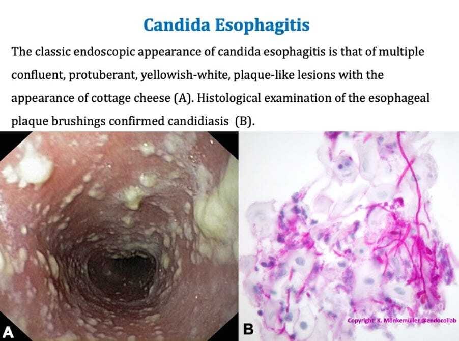
The classic endoscopic appearance of candida esophagitis is that of multiple confluent, protuberant, yellowish-white, plaque-like lesions with the appearance of cottage cheese.
Welcome to 5,991 motivated Endoscopists! Today we are going over Candida Esophagitis.

A 68-year-old man with a 10-year history of type 2 diabetes mellitus presented with dysphagia to solid foods for one week. He described the sensation as “food sticking" in the upper retrosternal area. Esophagogastroduodenoscopy revealed multiple confluent, protuberant, yellowish-white, plaque-like lesions with the appearance of cottage cheese covering the esophageal mucosa (Panel A). Histological examination of the esophageal plaque brushings confirmed candidiasis (Panel B). The patient was treated with oral fluconazole for 7 days with complete resolution of symptoms.
Candidiasis is a common opportunistic infection among immunocompromised patients usually involving the oropharynx, esophagus, and vagina. Patients with esophageal candidiasis typically present with dysphagia. An important distinction should be made between the 2 symptoms related to swallowing: odynophagia or painful swallowing is a complaint related to esophageal ulcer, whereas dysphagia or the sensation of “food getting stuck” is more commonly caused by esophageal strictures (benign or malignant) or candidiasis. Extensive esophageal candidiasis, however, can present with both symptoms. Endoscopic evaluation is the most sensitive method for diagnosis of Candida esophagitis. The characteristic white and yellow “cottage cheese” plaques are considered highly suggestive of candidiasis. In the appropriate clinical setting (e.g., immunosuppressed patient with thrush and dysphagia), a trial of empiric anti-fungal therapy with fluconazole can be attempted. If the condition fails to respond in less than a week, an endoscopic evaluation with biopsy and/or brushings should be carried out.

