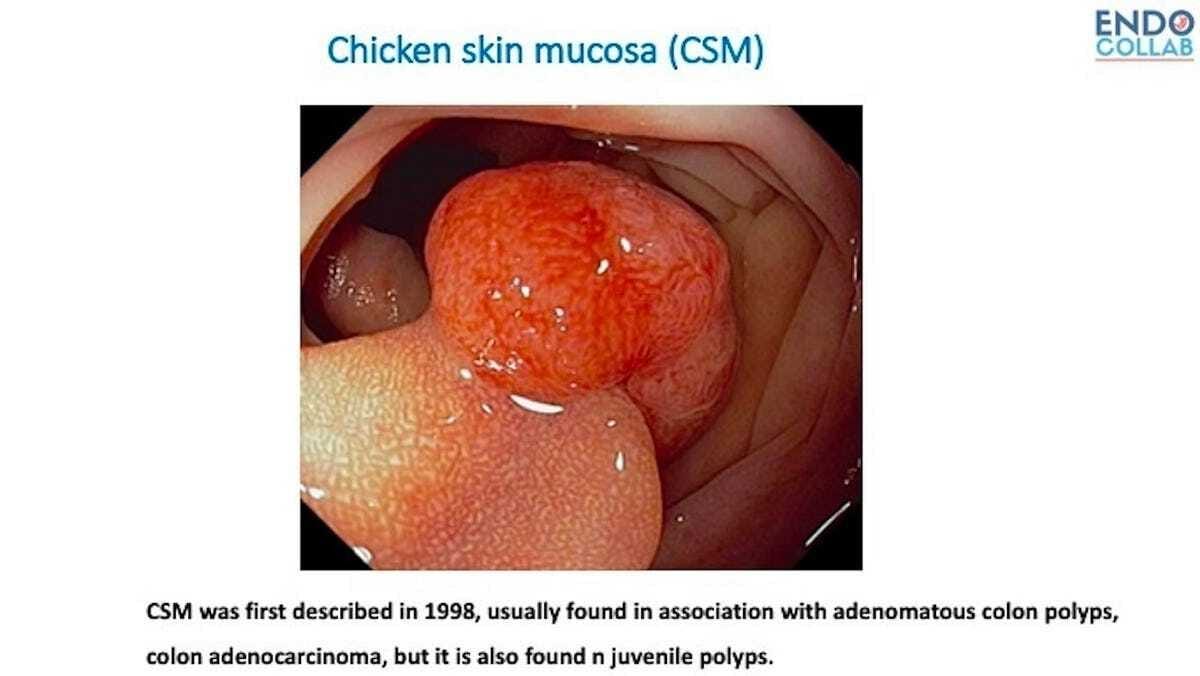
Chicken Skin Mucosa
Does the White Deposit (Chicken Skin Sign) Around the Base of the Polyps Have Any Prognostic Value?
“Chicken skin” mucosa (or “goose skin” in German-speaking countries, or “fish skin” in the Mediterranean and some Far East countries) was first described in Western countries in1998 by Shatz et al. (Am J Gastroenterol).
The authors found that CSM was associated frequently with adenomatous polyps and adenocarcinoma. Histologically, CSM is caused by an accumulation of lipid-laden macrophages. Subsequently, Nowicki et al (J Ped Gastroenterol Nutr, 2005) found that CSM is found in up to 75% of juvenile polyps. By using CD68 stain, which is specific for macrophages, the authors confirmed that CSM is due to lipid-laden macrophages and not associated with increased malignancy.
We had learned from Prof. Parra-Blanco that the first description of CSM was by Prof. Muto from Japan.
Prof. Parra-Blanco: “… this was described as “white spots” (hakuhan in Japanese) by Prof Tetsuichiro Muto in 1981 . But the name “chicken skin mucosa” is more popular in Western countries. It was described in Japanese, but this is the abstract in English.” https://www.jstage.jst.go.jp/article/gee1973b/23/2/23_2_241/_article/-char/en
Shatz BA, et al. Colonic chicken skin mucosa: an endoscopic and histological abnormality adjacent to colonic neoplasms. Am J Gastroenterol 1998;93:623–7.
Nowicki, M et al. Colonic Chicken-Skin Mucosa in Children with Polyps is not a Preneoplastic Lesion, Journal of Pediatric Gastroenterology and Nutrition. 2005;41:600-606.
[Link]


