Quick Tips and Tricks for Endoscopic Band Ligation of Esophageal Varices

Quick Tips and Tricks for Endoscopic Band Ligation of Esophageal Varices
By
Diana Dougherty, MD (Gastroenterology Fellow) and
Klaus Mönkemüller, MD, PhD, FASGE, FJGES
Professor of Medicine, Virginia Tech Carilion School of Medicine, Virginia, USA; Universidad de la República, Montevideo, Uruguay; University of Belgrade, Belgrade, Serbia, Universidad Espiritu Santo; Ecuador, University of Osijek, Croatia
A 58-year-old patient with cirrhosis (Child-Puch B, MELD 20) presented for EGD for screening esophageal varices. He was found to have large esophageal varices with red spots or markings (Figure 1 A) and four columns of varices (Figure 1B). Figures 1C and 1D show additional examples of red markings or spots (yellow arrows). These red spots are areas of wall thinning that may rupture and result in hemorrhage. The light blue arrow on Figure 1D shows a red marking, but here it is also possible to see some “bumps” or surface irregularity suggestive of significant wall thinning.

Figure 1
The key steps to follow when using band ligation for esophageal varices are:
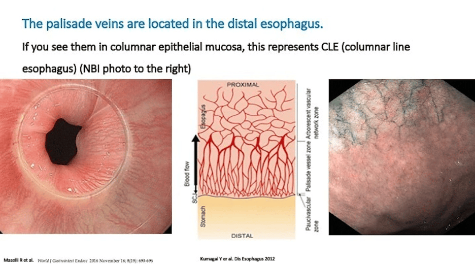
a) Determine the varix to be banded. This is accomplished by evaluating red markings or spots. In addition, banding should mainly be done to the distal 5 cm of esophagus, as it is extremely rare that varices in the mid-esophagus bleed. The highest-pressure gradients are at the GE junction and Z-line. Remember that the varices originate from the palisade veins located in the distal esophagus (see here https://community.endocollab.com/posts/25008135?utm_source=manual) (Figure 2, below)

Figure 2
b) Make sure that the stomach is empty, suction all remaining fluid and air before loading the banding device on the scope. Stomach contents induces retching and vomiting, and this may hinder endoscopy or even result in Mallory Weiss tears or bleeding.

Figure 3
c) The white strings of the cap with bands should align with the working channel of the scope (Figure 3A, 3B). In this case the working channel is on the left, around the 7 ‘clock position. In other scopes the working channel is on the 6 o’clock position. If the strings are not aligned with the working channel of the scope, there will be “crossing” the field and also impede adequate suction of the varix inside the cap.
d) Not infrequently, when introducing the scope with the banding cap, the interior of the cap will accumulate saliva, debris or lubricant gel (Figure 3A, above). A useful trick to get rid of this debris is to place the cap to a stomach mucosa (3B) and then flush with water (3C).
e) The clean cap is also useful for visualization of the esophageal varices, particularly the red markings or spots (Figure 3D).
f) Whenever suctioning on a varix, do it gently, with continuous suction, but do not suction in such a way that the entire cap is occluded by the varix. If too much tissue (varix) is strangulated by a band, partial esophageal obstruction may occur.
g) We recommend “gradual” suctioning. Figure 3E shows approx. one third of cap “filled” with varix. The ideal suction amount is shown in Figure 3F (two thirds of cap filled with varix). At that point it is important to release the band.
h) After the band has been released, no more suction should be applied. Indeed, soon after releasing the band we gently apply some air, and minimal amount of water (using the water pedal). Simultaneously, we pull the scope back a little bit. This combination of maneuvers (air, water and pulling the scope) “detaches” the suctioned varix (Figure 3G).
i) Careful inspection of the banded varix is important, but do not apply air or CO2 as the distention may result in band detachment.
j) We also look at the proximal variceal column to document flattening or disappearance of the column. Often, it is not necessary to deploy all six bands, as four or five bands may have resulted in adequate obliteration of the submucosal blood flow.
There are more tricks to manage esophageal and gastric varices. You can also watch short quick tip videos or full lectures on the topic on endocollab.com

