How I Do It: Endoscopic Resection of Zenker’s Diverticulum

How I Do It: Endoscopic Resection of Zenker’s Diverticulum
By Jay Bapaye, MD, Klaus Mönkemüller, MD, PhD, FASGE, FJGES
Virginia Tech Carilion School of Medicine, Virginia, USA
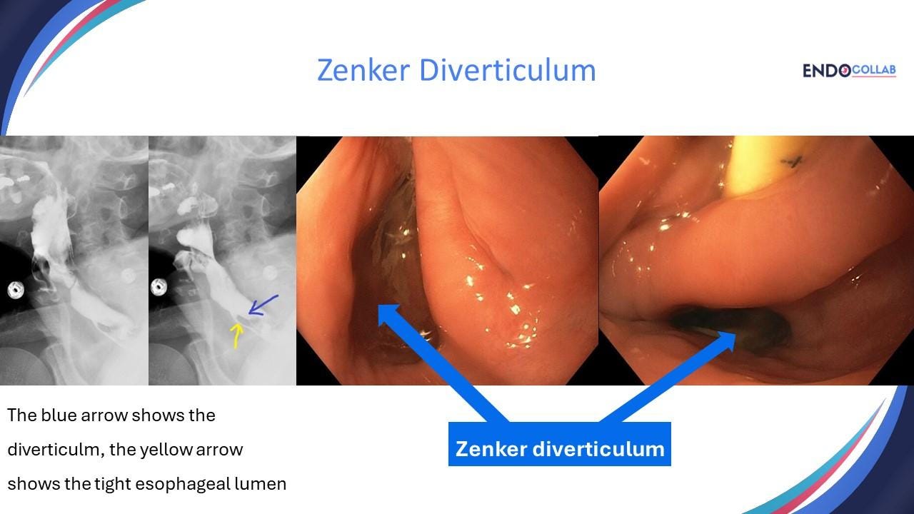
Zenker's diverticulum, also known as posterior pharyngeal pouch, is a pulsion diverticulum, resulting from posterior herniation of esophageal mucosa and submucosa into Killian's triangle, an area of least resistance situated above the upper esophageal sphincter (cricopharyngeal muscle) and below the inferior pharyngeal constrictor muscle (1-3).
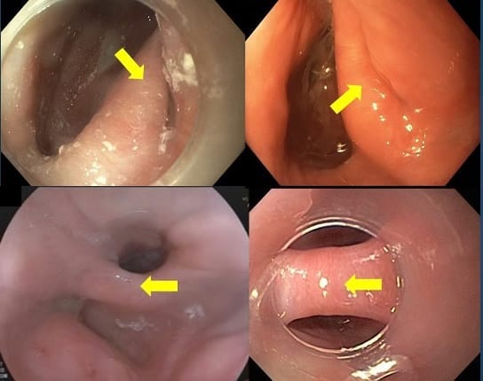
Figure 1. Spectrum of Zenker’s diverticulum. The yellow arrow points to the septum. Note that in some cases it is difficult to see the esophageal lumen, which appears as a “slid” (both upper photos).
Although open surgical techniques have historically been the criterion standard for treatment, endoscopic options have become increasingly used (3). Cricopharyngeal myotomy using flexible endoscopy is a safe and effective technique for the management of Zenker's diverticulum. Potential benefits of this approach include shorter operative times, shorter postoperative admissions, and earlier progression of diet (3).
The most common treatments for Zenker diverticulum are open surgical diverticulectomy with or without cricopharyngeal myotomy, rigid and flexible myotomy (1). The myotomy consist of incision of the “neck” of the diverticulum, starting with mucosa, then submucosa and, finally the muscle (“myotomy”).
There are many endoscopic approaches to “resect” a Zenker’s diverticulum. These include argon plasma coagulation (APC), incision with various types of needles (needle knife, hook knife, IT-knife), stapling device, endoscopic scissors, and so forth.
Although everyone talks about “resection”, this is not what is performed. Indeed, during flexible endoscopy the main approach “remove” the diverticulum is to perform a septotomy, incising the septum in between the diverticulum and esophagus, which contains the cricopharyngeal muscle. Indeed, the main goal of the treatment is to cut the cricopharyngeal muscle.
The myotomy consist of incision of the “neck” of the diverticulum, starting with mucosa, then submucosa and, finally the muscle (“myotomy”) (Figure 2). Figure 1 shows the septi of different Zenker diverticuli.
The essential aspects to perform an endoscopic resection are a) to have the tools, and b) follow specific steps (Figure 2).
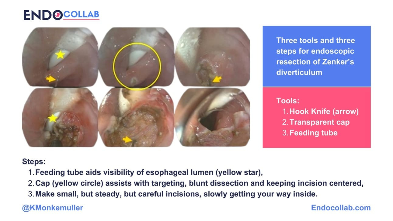
Figure 2. Zenker’s resection made easy using “the three tools and the three steps”.
The essential steps are:
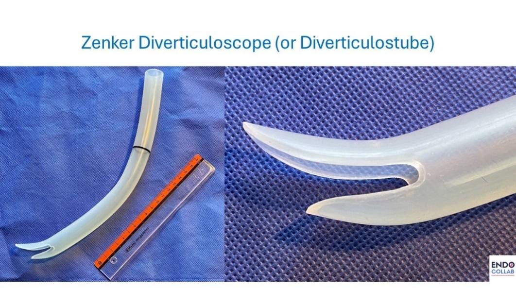
1.) Use a distal transparent cap on the endoscope (Figure 1 and Figure 2). The distal transparent cap stabilizes the endoscopic view, maintains a safe distance between the scope tip and the diverticular septum and allows for focused creating of the incisions using various devices. Additionally, the cap will allow for blunt dissection and, in case of bleeding, compression hemostasis. We have used the diverticuloscope in the past (Figure 3) but have found that it does not fit all Zenker diverticuli and thus use it only occasionally.
2.) Luminal vison: generally, the esophageal lumen and the diverticulum are clearly visible and distinct. However, more often than not, the septum is quite thick, and the esophageal lumen is barely visible (Figure 1). In this situation we like to place a wire (e.g. biliary guidewire) or a thin tube (e.g. Dobhoff or jejunal feeding tube) into the stomach, to maintain visibility of the esophagus and thus allow for controlled incision and dissection of the septum (Figure 2 and 4, panel D).
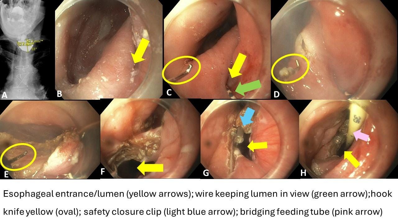
Figure 4. Endoscopic resection of Zenker’s diverticulum. The key steps to perform a safe septomyotomy in Zenker’s diverticulum are shown here.
3.) Incision and dissecting tool (utensil, device): Our preferred incision and dissection tool is the hook knife (Figure 4, yellow oval, panels C, D and E), as it permits hooking and cutting, as well as using the bend tip to dissect the muscle fibers one-by-one gradually and gently. A key aspect of septum dissection is to go step-by-step. First incise the mucosa, then the submucosa and lastly, muscle fibers, slowly, but steadily. Just below the cricopharyngeal muscle is a shiny, thin, film-like layer of the buccopharyngeal fascia which can be identified to ensure adequate myotomy.
4.) Safety closure. Once we have dissected down to the base of the septum, we prefer to place a closing clip at the apex of the dissection, as this is the most vulnerable area for perforation (and subsequent mediastinitis). There are many clips available, but we prefer to use one with a short stem (e.g. DuraClip), as this diminishes the chances of touching and rubbing the contralateral wall (Figure 4G and Figure 5).
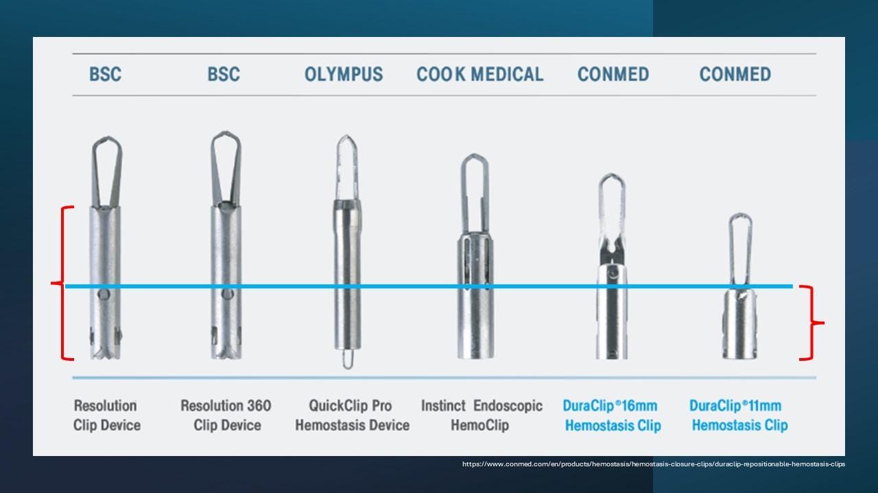
Figure 5. Comparison of various clips with focus on the length of the stem.
In sum, here we have presented a simple and safe endoscopic approach to resect Zenker’s diverticulum. This mini-review shows that there are a few essential steps and tools necessary to perform a Zenker’s resection, and these are modifiable, based on availability of tools and devices.
References:
Ferreira LE, Simmons DT, Baron TH. Zenker's diverticula: pathophysiology, clinical presentation, and flexible endoscopic management. Dis Esophagus. 2008;21(1):1-8.
Pescarus R., Shlomovitz E., Sharata A.M. Trans-oral cricomyotomy using a flexible endoscope: technique and clinical outcomes. Surg Endos. 2016;30:1784–1789.
Fan HS, Stavert B, Chan DL, Talbot ML. Management of Zenker's diverticulum using flexible endoscopy. VideoGIE. 2019 Jan 30;4(2):87-90. doi: 10.1016/j.vgie.2018.12.007. PMID: 30766952; PMCID: PMC6363821.
For more tips and tricks on diagnostic and therapeutic endoscopy visit endocollab.com
Caps in GI Endoscopy - In this case of Zenker's diverticulum
Endoscopic resection of Zenker's diverticulum made easy, safe and practical
No COI by JB or KM with any of the companies/utensils or products mentioned in this article.
Join the Premier Endoscopy Community - EndoCollab
Boost your endoscopy expertise with EndoCollab, the most comprehensive resource for endoscopists. As a member, you'll get:
• Access to 1000+ endoscopy strategies and techniques from top experts
• Ability to collaborate with a network of 1200+ endoscopists worldwide
• Case reviews to get answers to your toughest questions
• A vast library of endoscopy videos, images and courses at your fingertips
EndoCollab helps you stay at the forefront of endoscopy so you can provide the best care for your patients. Join our community of exceptional endoscopists today.
"EndoCollab is a game-changer for any endoscopist looking to expand their knowledge and connect with leading experts."
Don't miss this opportunity to transform your endoscopy practice. Become an EndoCollab member now.

Transform your practice with EndoCollab. Access a vast library of endoscopy techniques and join a network of experts. Elevate patient care – become part of our thriving community!
endocollab.com/join-endocollab

