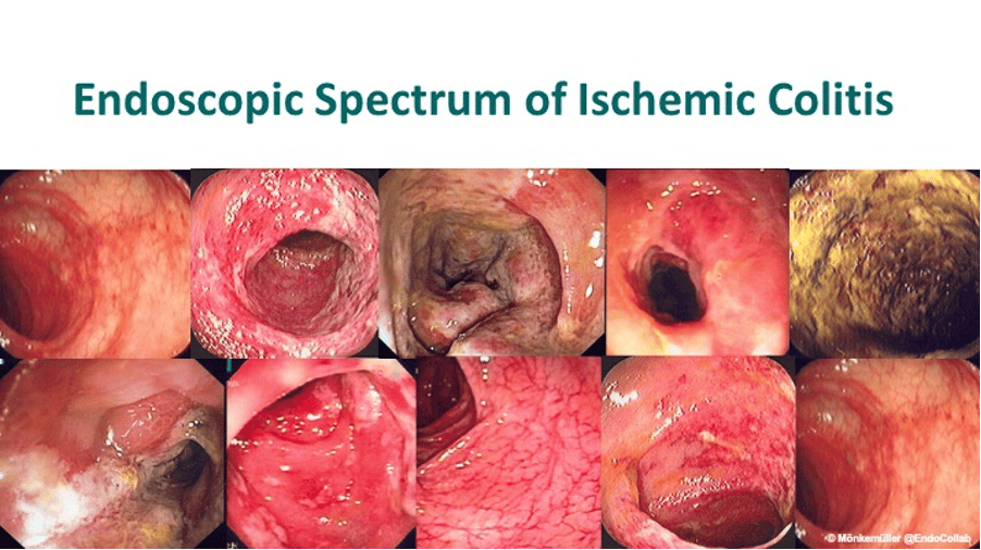
Endoscopic Spectrum of “Left”-Sided Ischemic Colitis

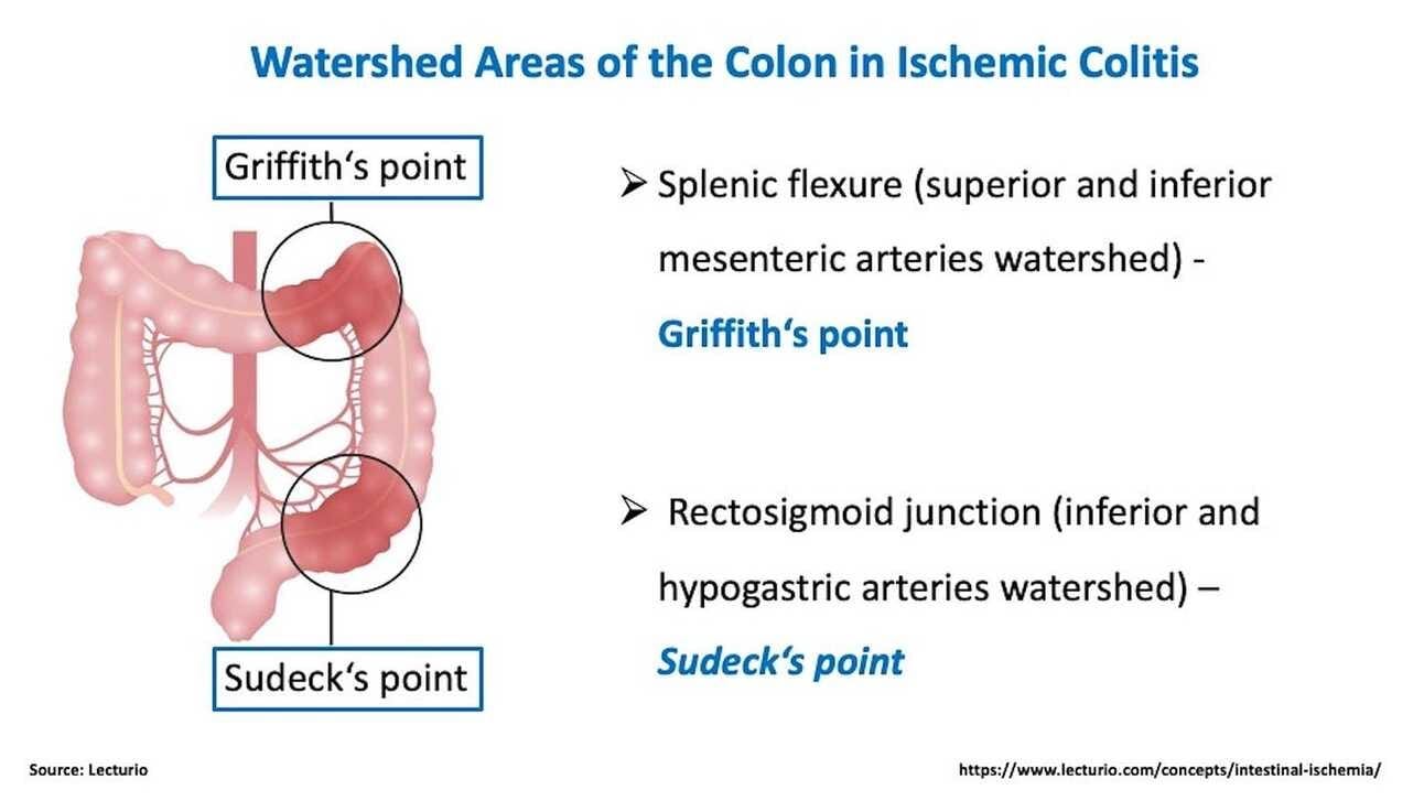
The endoscopic spectrum of ischemic colitis is broad. Key elements though are sparing of the rectum and segmental distribution, mainly in the left colon (at the watershed area, arc of Riolan).
Colonic ischemia occurs due to changes in systemic circulation and/or alterations in local mesenteric vasculature.
The most frequent areas affected are left colon (Griffith’s point), and superior rectum (Sudeck’s point), the lower rectum usually being spared because of its dual blood supply.
In mild ischemic colitis there are usually segmentally distributed patchy erythema, edema and subepithelial hemorrhages. In moderate colitis, in addition to changes seen in mild disease, there are localized erosions and ulcers, which may be confluent. Often, a linear ulcer in the mesenteric border of the colon is seen. This is known as colon single strip sign (CSSS) or Zuckerman’s sign. In severe colitis there are deep ulcers, luminal narrowing and strictures and frank necrosis.

More Ischemic Colitis on EndoCollab
A big thank you to this week's sponsors who help keep this newsletter free for the reader:
This vs That Digital Flashcards. Never be confused by similar GI diseases again! Flashcards for diseases that every endoscopist must know! Grab yours by clicking here.
EndoCollab. EndoCollab is an online community for GI endoscopists. With 800+ members, endoscopists discuss interesting cases, view on-demand video courses, and get new endoscopy tips every day. Join EndoCollab.

