Endoscopic Therapy for Bleeding Jejunal Diverticulum
by Hiral Patel, MD, Robert Moylan, MD and Klaus Klaus Mönkemüller, MD, PhD, FASGE, FJGES
Department of Gastroenterology, Carilion Memorial Hospital, Virginia Tech Carilion School of Medicine, Roanoke, USA
How Do You ACTUALLY Master GI Bleeding?
Want to see how a world-renowned expert approaches the toughest GI bleeds? Dr. Klaus Mönkemüller has edited a 224-page visual guide and atlas packed with hundreds of images, practical tips, and step-by-step case studies to show you how.
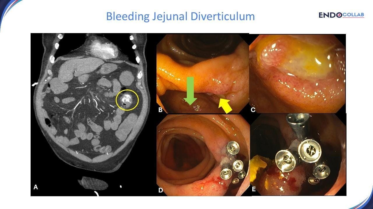
A 70-year-old man with history of hypertension and a stroke one year prior, since then using clopidrogel, presented with acute onset hematochezia and hemorrhagic shock. The patient was not taking any non-steroidals. A computed tomography angiography (CTA) showed active bleeding in the mid-jejunum (Figure 1 A). After hemodynamic stabilization an urgent single balloon enteroscopy was performed (Figure 1). Multiple small and large jejunal diverticula were found. There was a large diverticulum at about 100 cm distal to the duodenum with an ulcer on its inner wall (Figure 1C). The ulcer was treated with five clips. No further bleeding was observed.

Figure 1. Bleeding jejunal diverticulum. A. CTA showing active bleeding (extravasation of contrast, yellow circle) in the mid-jejunum. B. Large jejunal diverticulum (green arrow) with ulcer (yellow arrow). C. Large ulcer in the mouth of the diverticulum. D. Endoscopic hemostatic therapy using clips. E. Close up view of clipped ulcer.
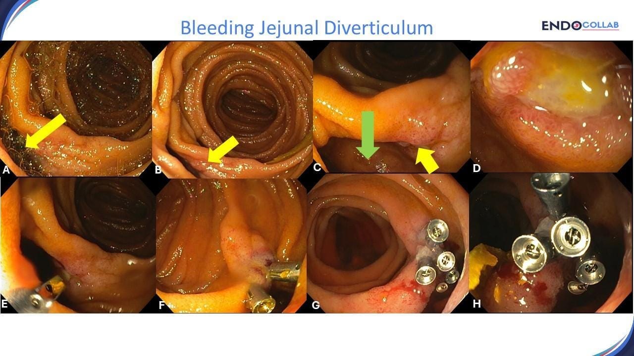
Figure 2 shows several tips and tricks on the endoscopic approach of jejunal diverticula. First, always look for different shapes of folds or darker areas behind or on the front of folds (A). These are clues for the presence of diverticula, blood or ulcers. Second, the use of simethicone will remove bubbles that disturb the view (B). Third, place the scope in a position that the worki g channel allws for targeting and treating the culprit lesion in a directed and easy manner (E). Fourth, use lots of clips to ensure the best possible closure or approximation of the ulcer and/ or hemostasis

Figure 2. Bleeding jejunal diverticulum. A. Dark contents inside of a jejunal diverticulum. B. irregular mucosa of the ulcerated mouth (entrance) of the diverticulum. C. Jejunal diverticulum (green arrow) with ulcer (yellow arrow). D. Large ulcer. E. The working channel of the scope on the left allowed for nice direction and placement of the clips. F. Two initial clips. G. and H. When confronted with a deep small bowel clip aggressive hemostasis is essential, i.e. better two additional clips than one to few”
In summary, this case is interesting for several reasons. First, a CTA was the first test performed for his hematochezia. CTA is being performed more often as a first line test in patients with gastrointestinal bleeding, especially if a “lower” (i.e. colonic) source is suspected (1). Second, after clinical stabilization we decided to perform a deep enteroscopy, obviating EGD and colonoscopy. This was a logical approach, as the source of bleeding was quite evident on CTA. Importantly, performing a capsule endoscopy may have delayed the treatment of the bleeding diverticulum. And finally, the concept of emergent deep enteroscopy proved to be of value, as the culprit lesion was found. Emergent double balloon was first described in 2009 and its use has been confirmed by various authors (2, 3). Finally, this report also describes key tips for reaching a diagnosis and achieving hemostasis in bleeding jejunal diverticulosis.
References:
Wu LM, Xu JR, Yin Y, Qu XH. Usefulness of CT angiography in diagnosing acute gastrointestinal bleeding: a meta-analysis. World J Gastroenterol. 2010 Aug 21;16(31):3957-63. doi: 10.3748/wjg.v16.i31.3957. PMID: 20712058; PMCID: PMC2923771.
Mönkemüller K, Neumann H, Meyer F, Kuhn R, Malfertheiner P, Fry LC. A retrospective analysis of emergency double-balloon enteroscopy for small-bowel bleeding. Endoscopy. 2009 Aug;41(8):715-7. doi: 10.1055/s-0029-1214974. Epub 2009 Aug 10. PMID: 19670141.
Pérez-Cuadrado Robles E, Bebia Conesa P, Esteban Delgado P, Zamora Nava LE, Martínez Andrés B, Rodrigo Agudo JL, López Higueras A, López Martin A, Latorre R, Soria F, Pérez-Cuadrado Martínez E. Emergency double-balloon enteroscopy combined with real-time viewing of capsule endoscopy: a feasible combined approach in acute overt-obscure gastrointestinal bleeding? Dig Endosc. 2015 Mar;27(3):338-44. doi: 10.1111/den.12384. Epub 2014 Nov 3. PMID: 25251991.
No COI by HP, RM or KM with any of the companies/utensils or products mentioned in this article.
Whenever you're ready, here are some ways EndoCollab can help you:
The Guide to GI Bleeding: Get our new #1 best-selling book. It's a 224-page visual atlas designed to help you master endoscopic hemostasis with hundreds of high-resolution images, case studies, and practical tips from world-renowned experts.
The EndoCollab Membership: Join our premium community for digestive disease professionals. Get lifetime, yearly, or monthly access to exclusive content, a growing library of video courses, and connect directly with experts and peers in our private members-only space.

