Esophageal Varix with Nipple Sign
Rami Musallam, MD, Gastroenterology Fellow,
Klaus Mönkemüller, MD, PhD, FASGE, FJGES, Professor of Medicine
Department of Gastroenterology, Carilion Memorial Hospital, Virginia Tech Carilion School of Medicine, Roanoke, USA
A 50-year-old patient with a history of cirrhosis due to ethanol use presented to the emergency room with hematemesis and melena for one day. The patient had clinical features consistent of chronic liver disease (spider angiomas, ascites, palmar erythema and jaundice). The hemoglobin was 8 gr/dl. An EGD was performed (Photo).

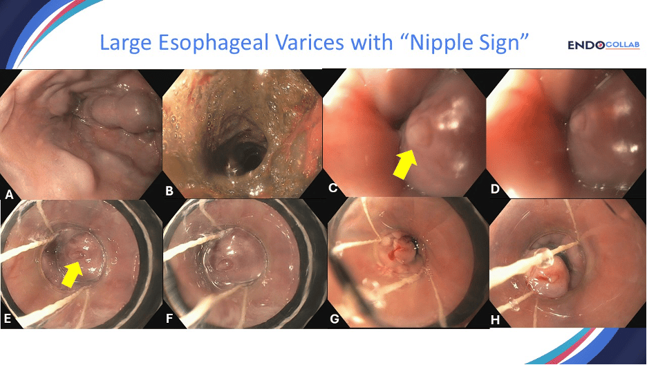
Figure 1. Large esophageal varices. A. Three columns of esophageal varices extending from the distal esophagus to the mid-esophagus. B. The stomach was full of coffee-ground like material, bile and blood clots. C. Classic nipple sign shown with yellow arrow. D. Nipple sign. E. The transparent banding cap allows for great visualization of the nipple sign (yellow arrow). F. No arrow on this photo to allow for “eye training” to detect nipple sign”. G. and H. Successful endoscopic band ligation of varix with high-risk stigmata (nipple sign).
There are various signs of impending or recent hemorrhage from esophageal varices. These include red stops, red whale sign, nipple-like sign and active bleeding (oozing or spurting) (1, 2). The “nipple-like projection” or “nipple sign” was described in 1996 (1). This lesion represents an opening varix covered by fibrin, indicating a recent bleed with spontaneous “hemostasis”. Any varix with nipple-like projection is at extremely high risk of (recurrent) bleeding, as this fibrin plug may fall off any time. Whenever this sign is encountered a quick decision to place endoscopic bands must be made. In this case we saw large esophageal varices (Panel A), then saw the lesion (Panels C and D). The stomach was full of old blood. As there was a clear culprit explaining the recent hemorrhage (fibrin nipple, panels C, D). Further cleaning of the stomach might have resulted in loss of the fibrin plug on the varix and catastrophic bleeding. Therefore, a quick decision was taken to place endoscopic band ligation on the high-risk lesion (panels E to H).
Recognizing a high-risk marker like the "nipple sign" can be the difference between routine banding and a catastrophic bleed. But what about the countless other challenging scenarios in GI bleeding?
For a comprehensive, practical approach to every situation, from varices to obscure bleeds, get your copy of our new book, 'The EndoCollab Guide for GI Bleeding'. https://amzn.to/40ugFRB
And to continuously sharpen your skills with practical tips, expert case discussions, and a global community of endoscopists, become a paid member of EndoCollab today. Don't just read the cases—be part of the conversation. https://endocollab.com/join-endocollab/
References:
Caldwell SH, Bickston SJ, Yoshida C, Morse J, Yeaton P. Nipple-like projection from a varix: a high-risk marker? Gastrointest Endosc. 1996 Nov;44(5):634-5. doi: 10.1016/s0016-5107(96)70035-4. PMID: 8934186.
Wasserman RD, Abel W, Monkemuller K, Yeaton P, Kesar V, Kesar V. Non-variceal Upper Gastrointestinal Bleeding and Its Endoscopic Management. Turk J Gastroenterol. 2024 May 20;35(8):599-608. doi: 10.5152/tjg.2024.23507. PMID: 39150279.
No COI by RM or KM with any of the companies/utensils or products mentioned in this article.
Article PDF Download

Newsletter - Esophageal Varices Nipple Sign.docx.pdf
225.30 KB • PDF File

