Hemostasis of Bleeding Duodenal Ulcer Using Injection Gold Probe
Diana Dougherty, MD, and Klaus Mönkemüller, MD, PhD, FASGE, FJGES
Department of Gastroenterology, Carilion Memorial Hospital, Virginia
Virginia Tech Carilion School of Medicine, Roanoke, USA
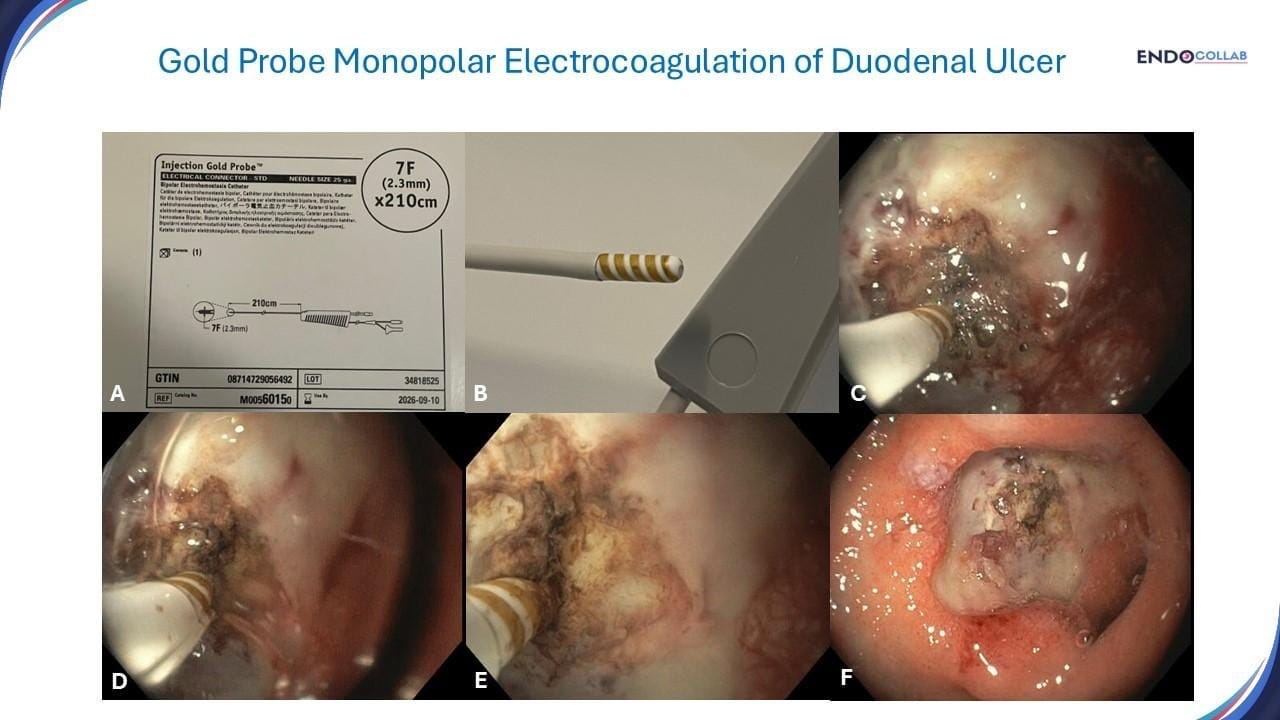
An 80-year-old man presented with melena. Esophagogastroduodenoscopy (EGD) revealed a large duodenal ulcer with a visible vessel, located anteriorly (Figure 1). Hemostasis was achieved with combination injection and compression electrocoagulation therapy using a injection gold probe.

Figure 1. Hemostasis with an injection gold probe catheter. A. Box of injection gold probe. The probe used was 7 Fr diameter. B. Notice the gold spiraled on the tip of the injection catheter. C. The gold probe can be placed laterally (tangentially) or perpendicular to the ulcer. D. The two important aspects are a) providing compression or pressure hemostasis and b) have enough gold touch the lesion to ensure cauterization of enough area (E). F. Successful hemostasis with disappearance of the visible vessel.
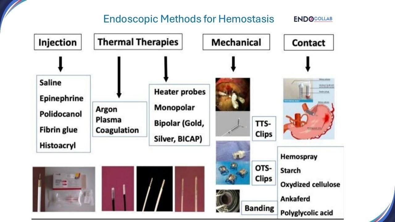
There is a large variety of tools to achieve endoscopic hemostasis. These are broadly classified into injection, thermal, mechanical and contact methods (1) (Figure 2).

Figure 2. Endoscopic Methods for Hemostasis. Thermal therapies are divided into argon plasma coagulation and probes that apply heat or electrosurgical current: monopolar, bipolar or multipolar electrocoagulation (MPEC) devices (1). With monopolar devices, the current passes through the patient and back to the unit via a return pad, whereas with bipolar or MPEC devices, the electric current is confined to the tissue between the electrodes within the instrument tip, obviating the need for a return pad (2).
An injection gold probe catheter is a bipolar hemostasis device that uses both injection and electricity to stop bleeding. We usually inject a mixture of saline epinephrine around the target area (e.g. visible vessel), followed by electrocauterization of tissue. The probe's tip heats tissue with electricity, which coagulates tissue and blood, thus stopping the bleeding. The probe's rounded tip provides uniform burn and coagulation. The probe's design reduces kinking, which helps it advance and tamponade tissue. The MPEC probe can be used tangentially to, or perpendicularly to, the bleeding source. Pressure is applied to compress and seal the walls of the bleeding vessel ("coaptive coagulation") (3). MPEC probes are available in 7F and 10F diameters with an irrigation port at the tip; the 10F probe requires the use of an endoscope with a 3.2 mm diameter instrument channel. Probe size, wattage, contact pressure and duration, and number of applications will vary depending on the lesion being treated (2).
Although we like to have this device in our toolbox for gastrointestinal bleeding, we don't use it very often, as there are other great options for hemostasis such as through-the-scope or over-the-scope clips. However, this case was ideal to use the injection gold probe as the ulcer was located anteriorly (i.e. on the working channel side of scope). If the ulcer is posteriorly, or on the right side of the scope, using injection or gold probe is quite difficult or impossible (3). In our experience we have not had any significant complications using the gold probe, but we have witnessed at least three cases of duodenal perforation in different hospitals. Therefore, we caution to be careful when performing pressure hemostasis, as the electrosurgical heat may damage deeper tissue levels (2, 4).
References:
1. Wasserman RD, Abel W, Monkemuller K, Yeaton P, Kesar V, Kesar V. Non-variceal Upper Gastrointestinal Bleeding and Its Endoscopic Management. Turk J Gastroenterol. 2024 May 20;35(8):599-608. doi: 10.5152/tjg.2024.23507. PMID: 39150279; PMCID: PMC11363156.
2. Parsi Me, et al. Devices for endoscopic hemostasis of nonvariceal GI bleeding (with videos). VideoGIE 2019;4:285-299.
3. Treating Upper Gastrointestinal Bleeding: An Update on Endoscopic Techniques - EndoCollab
4. Kumar VCS, Aloysius M, Aswath G. Adverse events associated with the gold probe and the injection gold probe devices used for endoscopic hemostasis: A MAUDE database analysis. World J Gastrointest Endosc. 2024 Jan 16;16(1):37-43. doi: 10.4253/wjge.v16.i1.37. PMID: 38313458; PMCID: PMC10835479.
Potential COI with the companies/utensils or products mentioned in this article: KM has been consultant for Ovesco, USA.
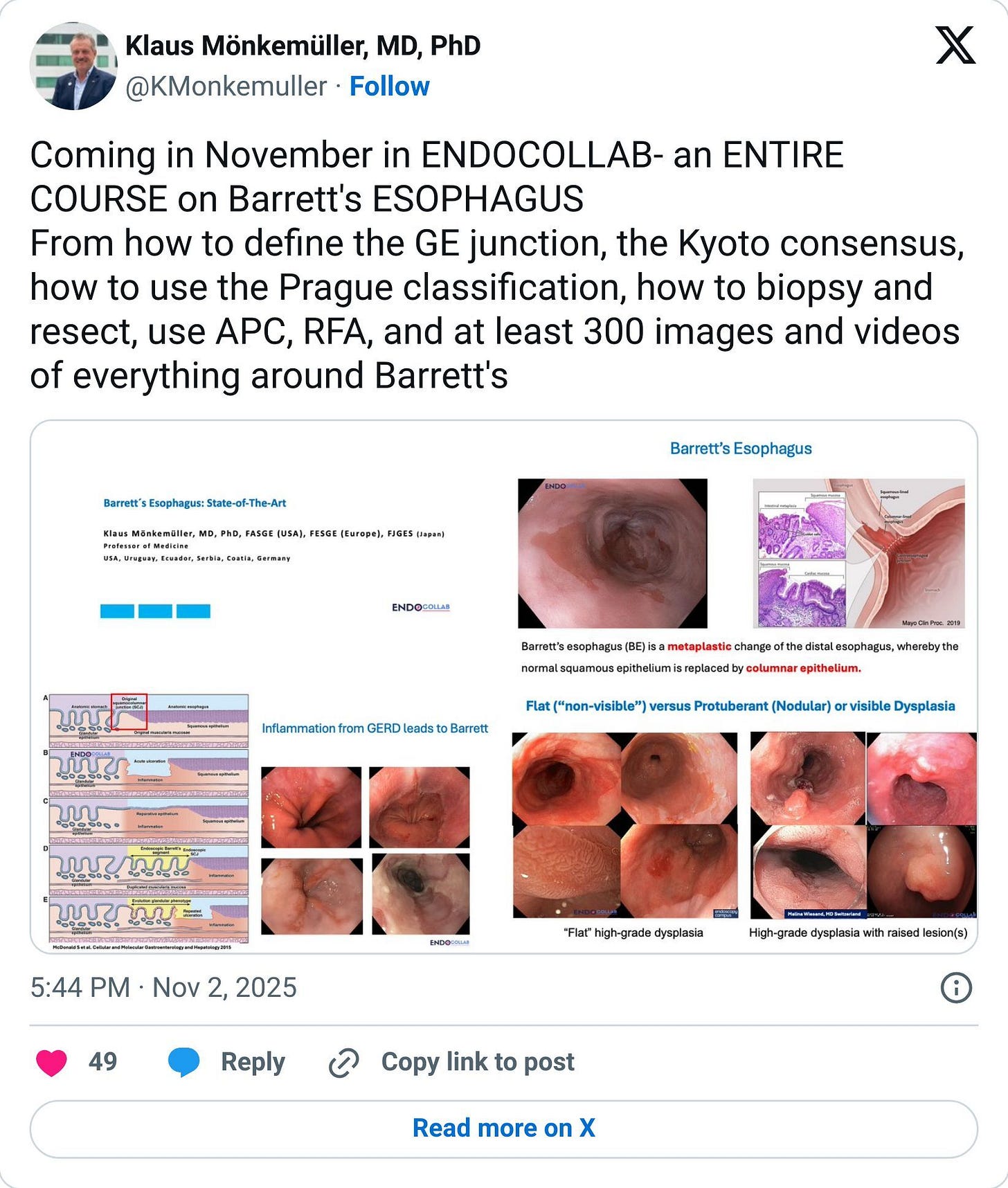
Coming in November in EndoCollab
An ENTIRE COURSE on Barrett's Esophagus
Join us this month as we dive deep into Barrett's Esophagus with comprehensive coverage including:
✓ How to define the GE junction
✓ The Kyoto consensus
✓ How to use the Prague classification
✓ Biopsy and resection techniques
✓ APC and RFA procedures
✓ At least 300 images and videos covering everything around Barrett's
Join EndoCollab today to access this comprehensive course and elevate your Barrett's management.

Through-the-scope clips have become the workhorse for many bleeding ulcers. But what about the tools we use less often, like the injection gold probe? This case is a perfect reminder of why a master endoscopist never forgets a tool in their kit.
This case shows a scenario where the gold probe offers excellent utility (effective "coaptive coagulation" on an anterior wall). Are you confident in your selection process for all hemostasis devices, even the ones you only pull out a few times a year?
📚 Get our new book: The EndoCollab Guide for GI Bleeding - Dive deep into strategies for acute GI bleeding, with practical frameworks for device selection and managing complications.
🔬 Join EndoCollab Premium: Become a member - Elevate your skills with our video library of annotated cases, technique demonstrations, and deep dives into every hemostasis device shown in this week's article.
Download the PDF of this article

newsletter_gold_probe.pdf
380.54 KB • PDF File

