Introducing “This vs That”, a Training Atlas to Improve Your Endoscopic Diagnostic Skills
A big thank you to this week's sponsors who help keep this newsletter free for the reader:
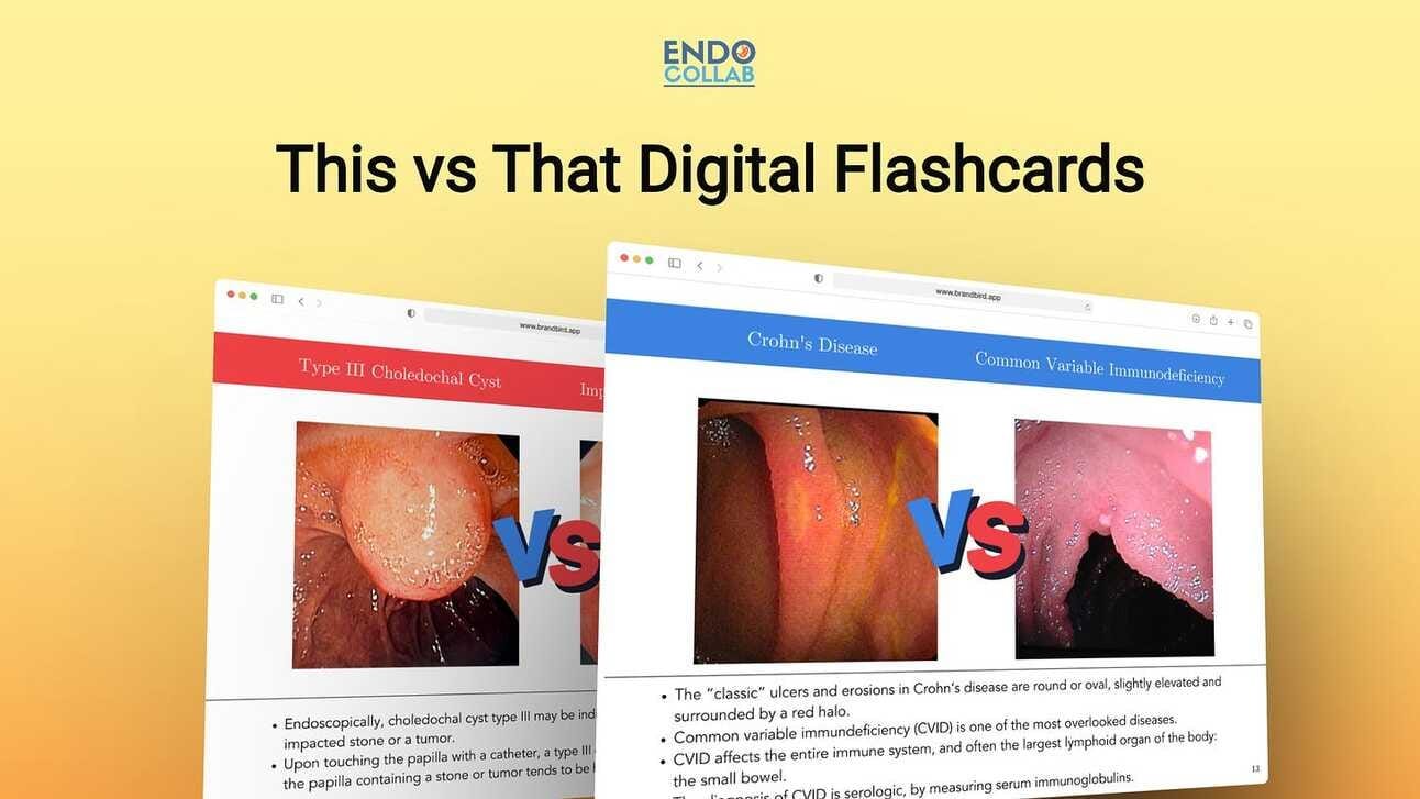
This vs That Digital Flashcards. Never be confused by similar GI diseases again! Flashcards for diseases that every endoscopist must know! Grab yours by clicking here.
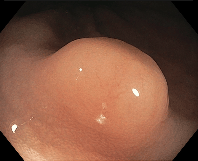
What Is the Diagnosis of This Rectal Polyp?

This is no simple polyp. And too often, there is a tendency to instinctively diagnose a “lipoma”. It is not.
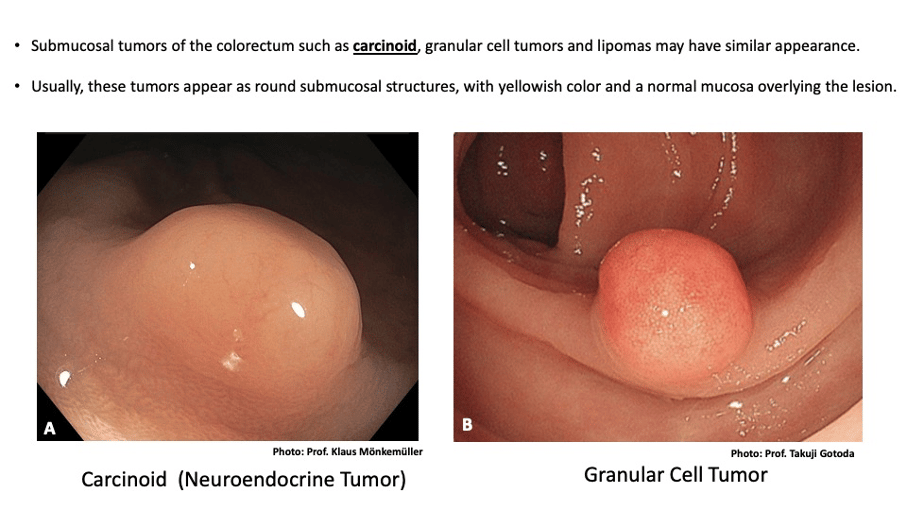
“This versus That”:

Whenever one sees a lesion like this (panel A) in the rectum or rectosigmoid area, it is a carcinoid (neuroendocrine tumor) until proven otherwise.
Note the round appearance and yellowish color with smooth mucosal pattern. Importantly, the pit pattern is Kudo type I, but the vascular pattern is Sano type 2, much different than that of a plain hyperplastic polyp. Why? Because carcinoid is a highly vascularized submucosal tumor.
Another possibility is a granular cell tumor (B), but these tumors are much less common than carcinoids.
Endoscopic ultrasound (EUS) may assist in evaluating the lesion.
Another great trick is injecting the submucosa. The lesion will not rise well. This is an important diagnostic step, as it will differentiate between mucosal and submucosal lesions.
In addition, submucosal injection will assist during the subsequent endoscopic resection of the lesion using either EMR or ESD techniques.
Never try to resect these lesions using plain snare polypectomy, as this technique often results in incomplete resection and stress for the patient and endoscopist, as the patient needs follow-up with resection of a scar with tumor remnant.
Get all 20 This vs That Flashcards by clicking here


