Technical Review: Endoscopic Resection of a Cardia Polyp Inside a Hiatal Hernia Sack Using the Duette Device
the Duette device was an ideal and safe tool to resect a polypoid lesion located in the cardia, inside a large hernia sack.

by Diana Dougherty, MD and Klaus Klaus Mönkemüller, MD, PhD, FASGE, FJGES
Department of Gastroenterology, Carilion Memorial Hospital, Virginia Tech Carilion School of Medicine, Roanoke, USA

Endoscopic Resection of a Cardia Polyp Inside a Hiatal Hernia Sack Using the Duette Device.pdf
216.02 KB • PDF File
Endoscopic Resection of a Cardia Polyp Inside a Hiatal Hernia Sack Using the Duette Device
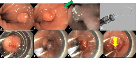
A middle-aged male patient presented for EGD because of dyspepsia and heartburn. On EGD he was found to have erosive reflux esophagitis grade C based on the Los Angeles classification, a 4 cm hiatal hernia and an incidental, sessile polyp inside of the hernia sack (Figures 1A-C). The polypoid lesion was covered with some exudate and the mucosa had a cerebroid pattern, with areas of erosions and spontaneous bleeding. There were no signs of fold convergence, scars, or loss of vascularity. A decision to resect the lesion was taken. We used the Duette device.

Why did we choose the Duette device in this situation?
First, the lesion was in a hernia sack and the sack was moving in and out during patient’s respiration. Second, the hernia sack allowed air (CO2) to constantly escape. And third, the patient had episodes of retching, as are often seen in patients with large hernias. Therefore, performing a traditional endoscopic mucosal resection (EMR) was less appealing, especially with a gastroscope having a working channel on the left side (about 7-8 o’clock position).
The advantages of performing the resection with the Duette system were the following: a) the lesion would be banded, and thus the mucosa to be resected is secured, as the endoscopist guides the resection based on were the band(s) is located, and b) the presence of a cap would allow for controlled resection and immediate suction (entrapment) and retrieval of the specimen.
The Duette device (Figure 1D) is essentially an endoscopic banding device for varices. The objective of suctioning and banding GI mucosal lesions is to a) reshape a flat or semi-sessile lesion into a “sessile” form, b) have the band at the base and assure a better endoscopic resection (R0).
Figure 1E shows the suctioning of the polyp and release of the band below the polyp.
The other novelty of the Duette device is the 5Fr hexagonal snare. If you would use a banding kit and then try to advance a traditional snare (usual diameter of the snare catheter or sheath is 7Fr), it would be impossible to push it through the working channel of the scope, as there is a string for the bands inside. When using Duette, pushing the 5Fr snare is quite easy. Figure 1F shows the snare being placed around the polyp. This hexagonal snare also has the advantage of having great expansile force and memory. It can be used to remove several lesions or pieces, such as large surface areas with dysplastic Barrett’s esophagus.
Depending on the amount of tissue caught, the snare can be placed below the band (if less tissue was caught), or above the band (if too much tissue was entrapped). In any event, the bands will not stay in place, and fall off immediately after the resection. But if too much tissue is caught and the snaring occurs below the band, there are higher chances of deep resection or even perforation. Therefore, it is important to observe the new post-suction-banded polyp to decide on the best strategy. In our case we placed two bands to safely secure the polyp while the patient was retching. Then we placed the snare between two bands (Figure 1G). The lesion was resected in its entirety, with a nice clean base (Figure 1H). Shortly after the resection, we advanced the scope into the stomach and “fired” all the bands and pulled the string out of the working channel of the scope. Then we went back to the hernia sack and suctioned the polyp into the cap and removed the polyp out of the patient (similarly to the removal of a “meat impaction” using cap).
Histology revealed an inflammatory polyp with intestinal metaplasia but no cancerous cells. The patient had an uneventful post-operative course and is on PPIs for the erosive reflux esophagitis.
In sum, the Duette device was an ideal and safe tool to resect a polypoid lesion located in the cardia, inside a large hernia sack.
No COI by DD or KM with any of the companies/utensils or products mentioned in this article.
Poll
In your practice, which method do you most commonly use for resecting polyps in challenging locations like a hiatal hernia sack?
Traditional endoscopic mucosal resection (EMR)
Duette device or similar banding-assisted resection
Endoscopic submucosal dissection (ESD)
Next Steps
Join EndoCollab: EndoCollab is your go-to platform for advancing your skills in gastroenterology and endoscopy. Gain access to exclusive content from top practitioners to enhance your practice.
Membership Features:
Video lessons on essential and advanced techniques.
Expert-led classes and practical tips.
Community-driven insights and peer discussions.
Join 1,200+ healthcare professionals taking their expertise to the next level.

