Tips and Tricks to Manage a Mallory Weiss Tear
The Practicing Endoscopist by EndoCollab

Tips and Tricks to Manage a Mallory Weiss Tear
by Diana Dougherty, MD and Klaus Mönkemüller, MD, PhD, FASGE, FJGES
Department of Gastroenterology, Carilion Memorial Hospital, Virginia Tech Carilion School of Medicine, Roanoke, USA
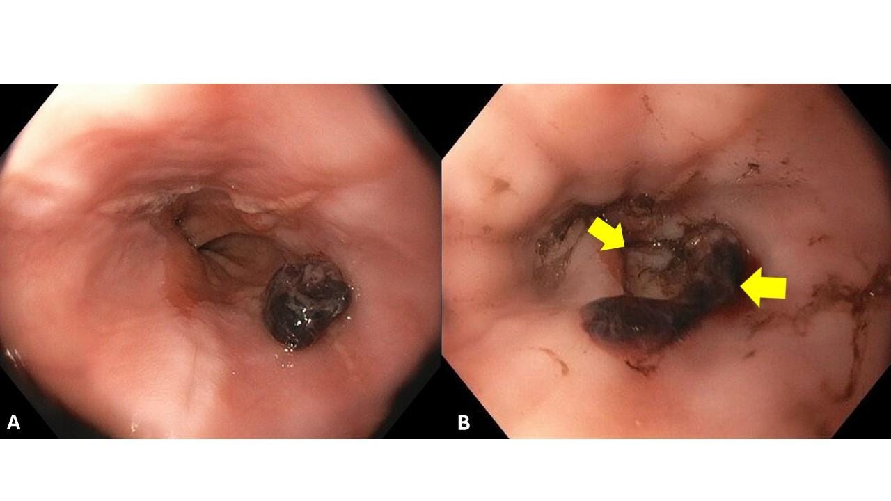
A young patient presented with massive upper gastrointestinal bleeding resulting in hypotension and requiring transfusion of four units of packed red blood cells. Once the patient was stabilized an emergent EGD was performed. A Mallory Weiss tear was diagnosed (Figure 1). An adherent blood clot was present, and there was active blood oozing from the tear. The size of the tear was estimated to be about 15 mm long.

The Key Initial Steps to Plan an Adequate Endoscopic Therapy for Mallory Weiss Tear Are To:
Determine its location (you want to plan potential utensils, such as clips, band, over-the-scope clip, injection, and the location of the working of the scope in relation to the lesion will assist you in determining what hemostatic method to use),
Analyze its morphology (length, depth, extension into stomach),
Make sure the stomach is as clean as possible, remove clots and blood, as stomach contents will decrease the endoscopic view, stimulate emesis and retching and may result in intra- or post-procedure aspiration.
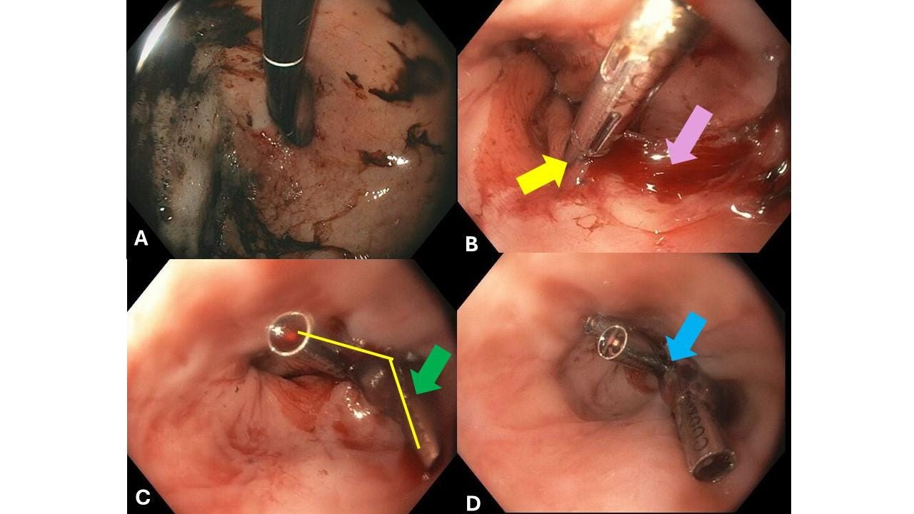
Perform a nice retroflexion and examine the cardia and any possible hiatal hernia (rule out any extension of the Mallory-Weiss tear into the stomach and rule out more tears or other lesions) (Figure 2A)
Endoscopic Therapy:
There are multiple endoscopic therapies used for achieving endoscopic hemostasis for Mallory-Weiss tears: injection of saline/epinephrin or glue, through-the-scope clips (hemoclips), over-the-scope clip, hemostatic powders, and gels (Hemospray, Purastat).

We decided to use clips for the because the Mallory Weiss lesion was large, there were lots of muscle fibers visible, indicating the tear had involved mucosa, muscularis mucosae, submucosa, and muscle (Figure 2B, pink arrow). By using clips, we would not only ensure hemostasis, but also closure of the wound, which cannot be achieved with topical or injected agents (powders, gels, glues). This patient had also been transfused with multiple units of blood, suggesting that deeper muscular vessels were involved by the tear. An important tip when using clips to close Mallory Weiss tears (or really, any large endoluminal GI defect) is to place the first clip on one of the edges of the tear (laceration or perforation) (Figure 2B, yellow arrow). In this case we chose to place the first clip distally, to prevent the laceration from extending into the cardia and stomach. Placing the first clip on the edges also helps create an initial closure and a mucosal “bump” which will facilitate application of other clips. We chose NOT to place the first clip proximally, as this would potentially impede or make placement of the next clips more difficult. The second clip was placed proximally, creating a “V” with the second clip (Figure C). This “V” configuration is allowed for a final placement of a third clip at the base of the “V”. successfully closing the laceration and achieving hemostasis. Figure D shows adequate tissue apposition.
In sum, Mallory Weiss tears can result in catastrophic bleeding. Endoscopic therapy should be aimed at both hemostasis and closure of the defect, especially in lesions that are deep and result in significant bleeding. Various endoscopic therapies are available, of which hemoclips are well-suited to achieve both hemostasis and tissue apposition (i.e. closure of defect).
No COI by DD or KM with any of the companies/utensils or products mentioned in this article.

Transform your practice with EndoCollab. Access a vast library of endoscopy techniques and join a network of experts. Elevate patient care – become part of our thriving community!
endocollab.com

