Tips for Management of Colon Polyps with Thick and/or Long Stalks Using Endoloop
By Varun Kesar, MD and Klaus Mönkemüller, MD, PhD, FASGE, FJGES
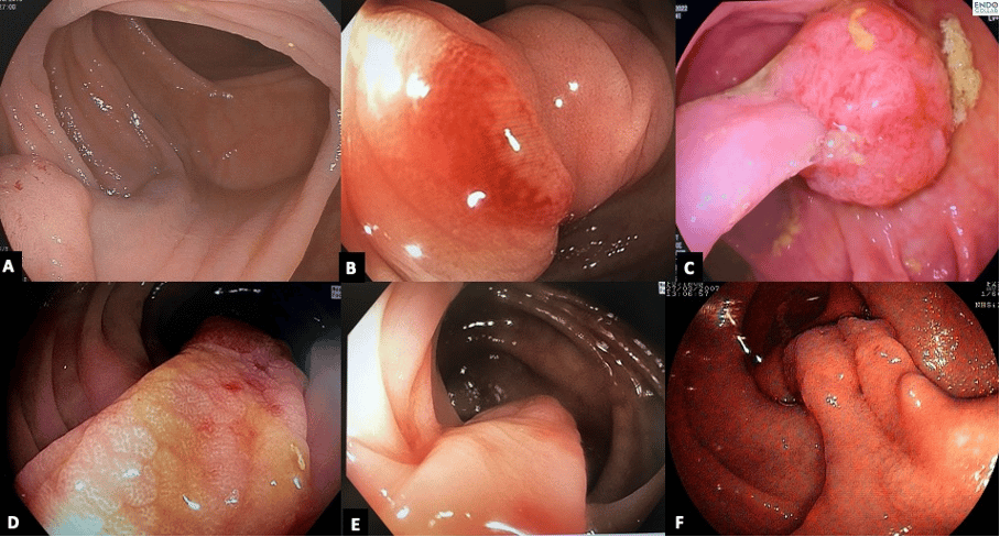
Pedunculated polyps (Paris 0-Ip) with thick and long stalk (Figure 1) pose a resection challenge as the pedicle usually has one or more big arteries and thus, the risk of intra- or post-polypectomy bleeding is increased. Therefore, pre-resection or post-resection interventions to prevent post-polypectomy bleeding are mandatory. These include clipping, injection, using endoloop or a combination of some of them.

In dealing with 0-Ip lesions, our approach is pre-resection endoloop placement. While endoloops may resemble snares, their endoscopic handling is different because they can't be opened and closed as easily. We pass the catheter with the endoloop proximal to the polyp head, slowly expose the loop, and then carefully place it on the polyp head.
In the 0-Ip lesions shown here we proceeded with pre-resection endoloop placement. Although endoloops look like a snare, endoscopic handling is different, as these cannot be opened and closed at will (Figure 2A). Thus, we like to pass the catheter with the endoloop proximal to the polyp head (Figure 2B), expose (“open”) the loop slowly and then pull scope with the opened loop proximally, slowly allowing for the loop to place itself on the polyp head. Once the endoloop can be clearly seen “under” the polyp head and around the stalk we wiggle the scope and or the loop-catheter to go as proximal to the stalk base. We like to tighten and place the loop about 10 mm above the base (Figure 2C).

When the assistant tightens the loop using the “pushing ring or band”, the endoscopist must gently, and minimally push the scope towards the stalk, staying close to the “field of action”. After tightening of the loop with the pushing band has occurred, the endoscopist starts pulling catheter through the working channel of the scope, using the right hand, while the assistant does the final release of the loop, pushing it out. At that moment, the long tail of the attached loop becomes visible and the polyp head starts changing color, acquiring a bluish discoloration (Figure 3A). Once the polyp has a purplish color we proceed with endoscopic resection in three steps: a) closing the snare and holding tight for about 30 seconds (this induces physiologic coagulation); b) applying a short burst of coagulation current; and c) applying cutting current (here we prefer the computer-controlled Endocut current) (Figure 3B). The resection process is the same when using clips, except that careful attention must be paid not to capture any the loop with the electrosurgical snare. Figure 3C shows on the importance of cutting well below the neoplastic tissue, to ensure complete resection (R0). Also, notice how cutting well below the polyp head, also ensure resection of neoplastic tissue extending into the stalk (Figure 3C).

Images by EndoCollab. See endocollab.com for more information, including videos, quick tips and lectures on these and many other practical endoscopy tricks.
Endoloop on EndoCollab
Tips for Management of Colon Polyps with Thick and/or Long Stalks Using Endoloop
Answer to Colon Polypectomy: What additional tools/techniques 🧰 would you use to resect this polyp?
Today's Quick Tip Video - Using Endoloop: a Step-by-Step Guide
Join EndoCollab
✅ 1,000+ endoscopy strategies
✅ 1,000+ endoscopists
✅ EndoCollab community to ask your questions
✅ No obligations, no contracts, CANCEL ANYTIME


